- Anatomical terminology
- Skeletal system
- Joints
- Muscles
- Heart
- Blood vessels
- Blood vessels of systemic circulation
- Aorta
- Blood vessels of head and neck
- Arteries of head and neck
- Veins of head and neck
- Blood vessels of upper limb
- Blood vessels of thorax
- Blood vessels of abdomen
- Blood vessels of pelvis and lower limb
- Blood vessels of systemic circulation
- Nervous system
- Respiratory system
- Digestive system
- Lymphatic system
- Female reproductive system
- Male reproductive system
- Endocrine glands
- Eye
- Ear
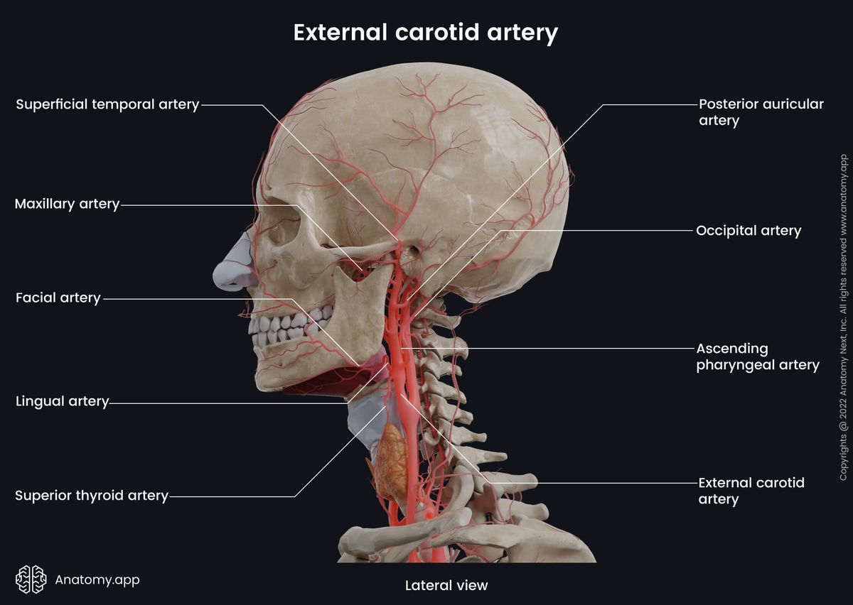
External carotid artery
The external carotid artery (Latin: arteria carotis externa) is a major blood vessel that arises from the common carotid artery and is located in the anterior neck triangle. It is one of two terminal branches of the common carotid artery. Every person has two external carotid arteries - one on each side of the neck. Overall, the external carotid artery and its branches supply arterial blood to the neck, face and scalp.
External carotid artery origin and course
The external carotid artery originates in the neck, arising from the bifurcation of the common carotid artery at the level of the superior border of the thyroid cartilage (C3 - C5 level). It happens within the carotid neck triangle. From the bifurcation, both carotid arteries ascend towards the external cranial base. The internal carotid enters the cranial cavity via the external opening of the carotid canal. In contrast, the external carotid artery ascends through the neck within the carotid sheath posterior to the ramus of the mandible and anterior to the external ear. The proximal portion of the external carotid is positioned anteromedial to the internal carotid artery.
At first, the external carotid artery curves slightly anteriorly. But, as it travels towards the mandibular neck, it inclines posteriorly. The proximal segment of the external carotid goes between the mandibular angle and the mastoid process of the temporal bone. On its course, the external carotid artery is crossed by the stylohyoid muscle and the posterior belly of the digastric muscle. Finally, the external carotid artery enters the parotid gland behind the neck of the mandible and divides into two terminal branches - the superficial temporal artery and the maxillary artery.
External carotid artery branches
On its course, the external carotid artery gives off the following six side branches:
- Superior thyroid artery - originates from the anterior surface;
- Lingual artery - branches off from the anterior aspect;
- Facial artery - arises from the anterior side;
- Ascending pharyngeal artery - derives from the medial surface;
- Occipital artery - stems from the posterior side;
- Posterior auricular artery - branches off from the posterior aspect.
As mentioned previously, the external carotid artery ends within the parotid gland by dividing into two terminal branches - the superficial temporal artery and the maxillary artery.
Superior thyroid artery
The superior thyroid artery arises from the anterior aspect of the external carotid artery at the level of the hyoid bone. It travels downward along the lateral margin of the thyrohyoid muscle to reach the upper pole of the thyroid gland.
The superior thyroid artery has various small branches that supply the thyroid gland and its adjacent soft tissue (some of the muscles of the larynx and muscles of the neck). The superior thyroid artery gives the following branches:
- Glandular arteries
- Infrahyoid artery
- Sternocleidomastoid artery
- Cricothyroid artery
- Superior laryngeal artery
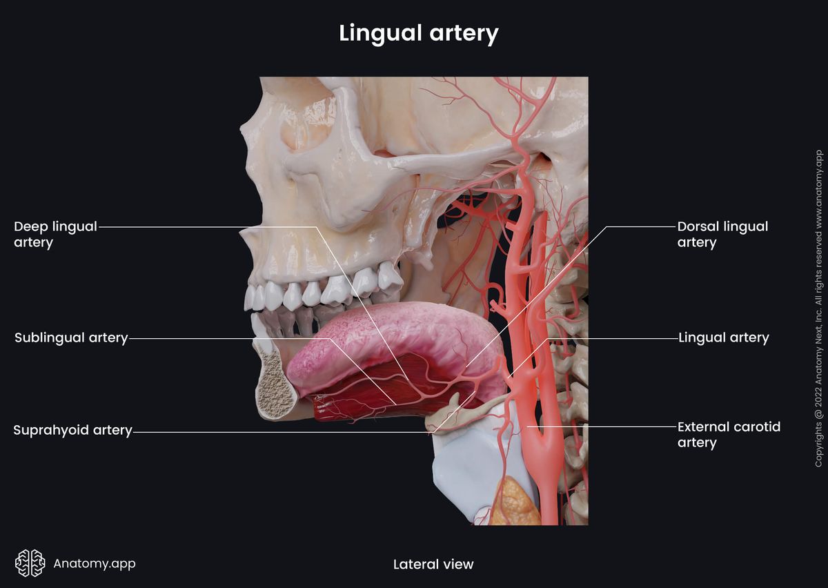
Lingual artery
The lingual artery also arises from the anterior surface of the external carotid artery at the level of the greater horn of the hyoid bone (C3 vertebra). At first, it travels medially within the carotid neck triangle. Then it forms a loop and courses anteroinferior. The loop is crossed by the hypoglossal nerve (CN XII). Further, the lingual artery goes along the superior aspect of the hyoid bone, reaches the posterior border of the hyoglossus muscle and passes forward between it and the middle pharyngeal constrictor.
As it reaches the anterior border of the hyoglossus muscle, the lingual artery ascends further anteriorly on the inferior surface of the tongue, going between the genioglossus and inferior longitudinal tongue muscle. Finally, it terminates by giving a deep lingual artery that reaches the tip of the tongue.
Overall, the lingual artery provides arterial blood supply to the tongue, soft palate, palatine tonsils, hyoid bone, sublingual gland and the floor of the mouth. Along its course, the lingual artery gives the following branches :
- Suprahyoid artery
- Dorsal lingual artery
- Sublingual artery
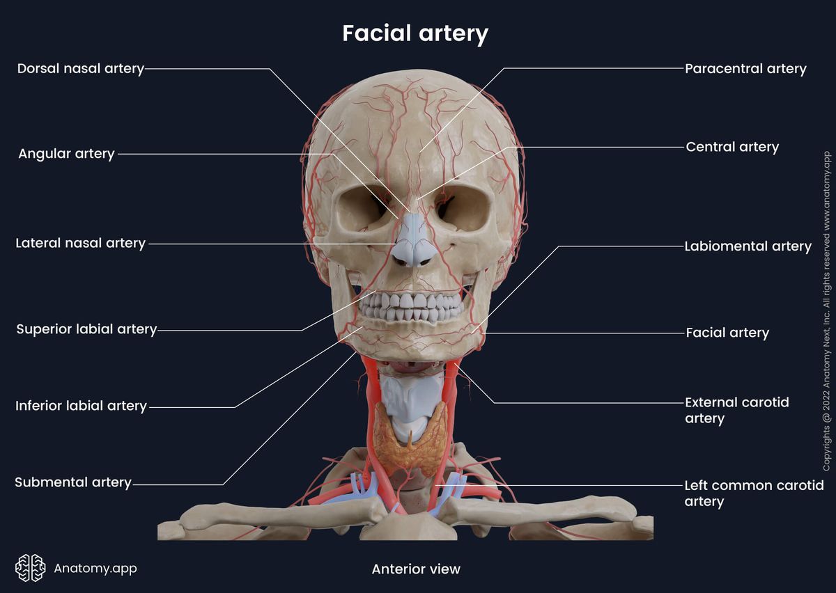
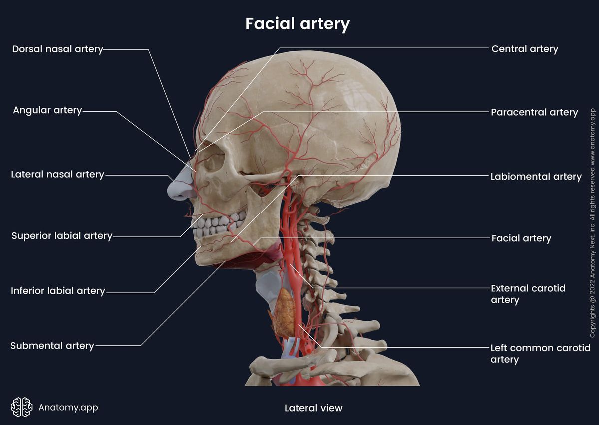
Facial artery
The facial artery originates above the lingual artery from the anterior aspect of the external carotid artery at the level of the mandibular angle. It travels in an anterosuperior direction, curves around the inferior border of the mandibular base and emerges on the face.
Further, it ascends across the mandibular body towards the angle of the mouth. From here, the facial artery passes upward along the lateral side of the nose to the medial canthus of the eye. The facial artery ends as the angular artery, which forms an anastomosis with the dorsal nasal artery of the ophthalmic artery at the medial angle of the orbit.
On its course, the facial artery gives off several branches, including the following:
- Ascending palatine artery - supplies the soft palate, Eustachian tube and palatine tonsil;
- Tonsillar artery - provides supply to the palatine tonsil;
- Submental artery - supplies the soft tissues of the submental area;
- Glandular arteries - provides supply to the submandibular gland and adjacent lymph nodes;
- Inferior labial artery - supplies the lower lip;
- Superior labial artery - provides supply to the upper lip, nasal septum and ala of the nose;
- Lateral nasal artery - supplies the dorsum and ala of the nose.
Ascending pharyngeal artery
The ascending pharyngeal artery is the only branch that arises from the medial side of the external carotid artery between the pharynx and the internal carotid artery. It is long and thin, and at the same time, it is the smallest branch of the external carotid artery.
The ascending pharyngeal artery travels along the posterolateral wall of the pharynx, going anterior to the longus capitis muscle and medially to the styloglossus and stylopharyngeus muscles. It originates near the carotid bifurcation and terminates by anastomosing with the ascending palatine branch of the facial artery and the ascending cervical branch of the vertebral artery.
The ascending pharyngeal artery has a relatively short common trunk before it divides into two major trunks - the neuromeningeal (intracranial) and pharyngeal (extracranial). Overall, the ascending pharyngeal artery with its branches supplies the following structures:
- Pharynx
- Soft palate and palatine tonsils
- Meninges of the posterior cranial fossa
- Tympanic cavity
- Eustachian tube
- Neck muscles (longus colli and longus capitis)
- Glossopharyngeal nerve (CN IX)
- Vagus nerve (CN X)
- Hypoglossal nerve (CN XII)
- Sympathetic trunk
- Some of the cervical lymph nodes
Occipital artery
The occipital artery originates from the posterior surface of the external carotid artery. It travels posterior and superior towards the mastoid process of the temporal bone, going parallel to the posterior belly of the digastric muscle. On its course, the occipital artery crosses the internal jugular vein, internal carotid artery, vagus nerve (CN X), accessory nerve (CN XI) and hypoglossal nerve (CN XII).
When reaching the mastoid part of the temporal bone, the occipital artery is accompanied by the greater occipital nerve, and both go within the occipital groove. Then the artery pierces the deep cervical fascia between the trapezius and sternocleidomastoid muscles and reaches the scalp, where it ascends within its superficial fascia.
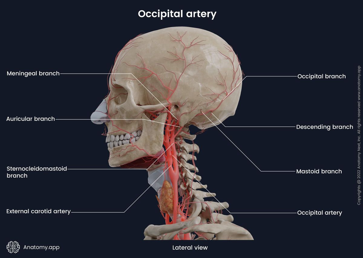
Along its course, the occipital artery gives the following branches:
- Sternocleidomastoid branch
- Auricular branch
- Mastoid branch
- Descending branch
- Occipital branch
- Meningeal branch
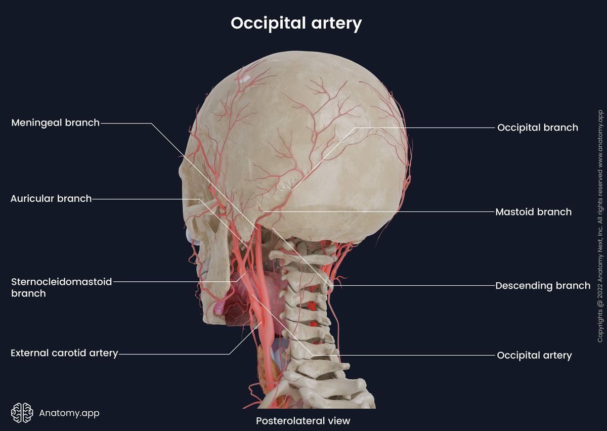
Overall, the occipital artery supplies the scalp of the occipital area, occipitofrontalis muscle, sternocleidomastoid, trapezius, some of the deep back muscles, auricle and the dura mater of the posterior cranial fossa.
Posterior auricular artery
The posterior auricular artery arises from the posterior aspect of the external carotid artery at the level of the superior border of the posterior belly of the digastric muscle. It ascends posteriorly towards the external ear, traveling between the styloid process of the temporal bone and the parotid gland.
When it reaches the auricle of the external ear, the posterior auricular artery passes in a groove between the auricular cartilage and the mastoid process of the temporal bone. Finally, the posterior auricular artery terminates within the scalp posterior to the auricle.
Along its course, the posterior auricular artery gives the following branches:
- Auricular branches
- Posterior tympanic branch
- Occipital branches
- Parotid branch
- Stylomastoid artery
Overall, the posterior auricular artery supplies adjacent neck muscles (digastric, stylohyoid and sternocleidomastoid), auricle, external acoustic meatus, tympanic cavity, semicircular canals, facial nerve (CN VII), mastoid antrum, mastoid air cells and the parotid gland.
Superficial temporal artery
The superficial temporal artery is the smallest of both terminal branches, supplying the forehead, temporal and parietal regions of the scalp. It arises from the external carotid artery within the parotid gland behind the mandibular neck.
The superficial temporal artery further ascends towards the scalp anterior to the ear, crossing over the zygomatic process of the temporal bone. It terminates above the zygomatic process at the level of the supraorbital margin by giving two terminal branches - frontal (anterior) and parietal (posterior). Besides the terminal branches, the superficial temporal artery gives the following branches:
- Parotid branch
- Transverse facial artery
- Anterior auricular branches
- Zygomatico-orbital artery
- Middle temporal artery

Overall, all these branches provide arterial blood supply to the external ear, parotid gland, lateral surface of the face, facial muscles and skin, temporalis and masseter muscles, as well as the temporomandibular joint.
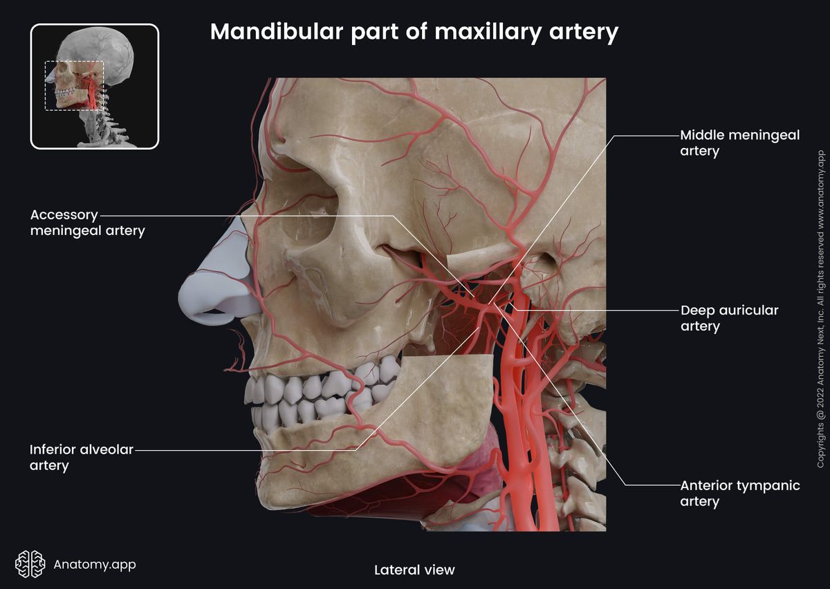
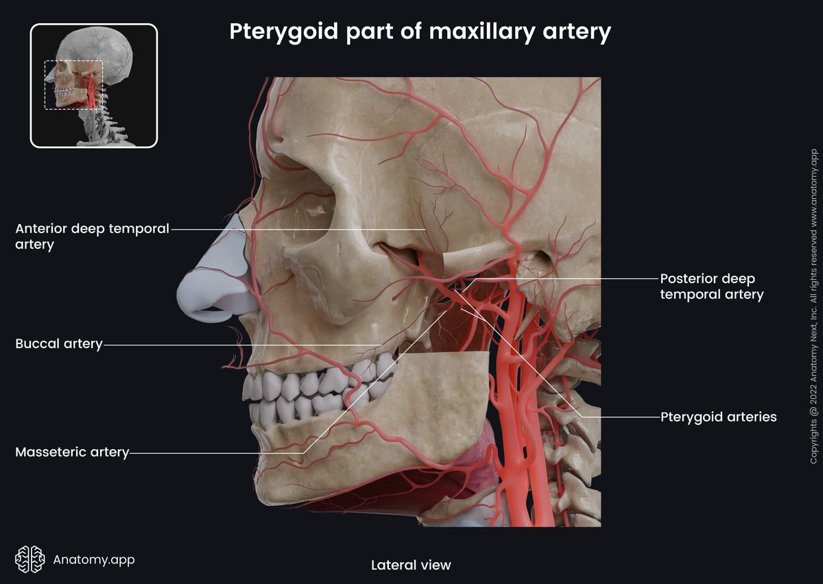
Maxillary artery
The maxillary artery is the largest terminal branch of the external carotid artery, and it also arises within the parotid gland. From the parotid gland, the maxillary artery courses anteriorly towards the pterygopalatine fossa, going between the sphenomandibular ligament and the neck of the mandible. At first, it travels through the infratemporal fossa and then enters the pterygopalatine fossa via the pterygomaxillary fissure.
The maxillary artery is divided into three portions based on its location:
- The first portion is the mandibular part, which is positioned between the mandibular neck and the sphenomandibular ligament.
- The second portion is the pterygoid part found lateral to the lateral pterygoid muscle.
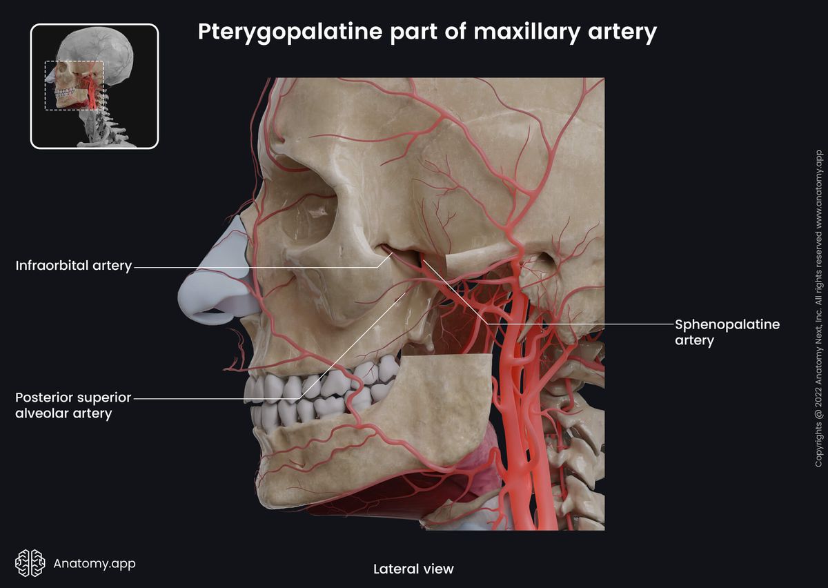
- And finally, the third portion is the pterygopalatine part. It is defined as the segment of the maxillary artery which passes through the pterygopalatine fossa.
Overall, the maxillary artery supplies the structures found within the infratemporal fossa, structures of the ear, palate, maxilla, mandible, teeth, gingiva, temporomandibular joint, nasal cavity, muscles of mastication and dura mater of the cranial cavity.
Maxillary artery branches
Each part of the maxillary artery gives off several branches. From the mandibular part of the maxillary artery arise the following arteries:
- Middle meningeal artery
- Inferior alveolar artery
- Deep auricular artery
- Anterior tympanic artery
- Accessory meningeal artery
The pterygoid part of the maxillary artery gives rise to the following branches:
- Anterior and posterior deep temporal arteries
- Masseteric artery
- Buccal artery
- Pterygoid arteries
And finally, the pterygopalatine part of the maxillary artery provides the following branches:
- Posterior superior alveolar artery
- Infraorbital artery
- Sphenopalatine artery
- Descending palatine artery
- Pharyngeal artery
- Artery of the pterygoid canal
Related structures of external carotid artery
Anterior to the external carotid artery is positioned the sternocleidomastoid muscle, platysma, deep cervical fascia, superficial cervical fascia and skin of the neck.
Posterior to the artery lies the superior laryngeal nerve, styloglossus and stylopharyngeus muscles, glossopharyngeal nerve (CN IX), pharyngeal branch of the vagus nerve (CN X) and the parotid gland.
Medial to the external carotid artery can be found the hyoid bone, pharyngeal wall, superior laryngeal nerve and the parotid gland. Lateral to the artery is the internal carotid artery.
References:
- Cramer, G. D. (2014). Clinical Anatomy of the Spine, Spinal Cord and Ans (Third Edition). Elsevier.
- Drake, R., Vogl, W., & Mitchell, A. (2019). Gray’s Anatomy for Students: With Student Consult Online Access (4th ed.). Elsevier.
- Gray, H., & Carter, H. (2021). Gray’s Anatomy (Leatherbound Classics) (Leatherbound Classic Collection) by F.R.S. Henry Gray (2011) Leather Bound (2010th Edition). Barnes & Noble.
- Mauro, M. A., Murphy, K., Thomson, K. R., Venbrux, A. C., & Morgan, R. A. (2021). Image-guided interventions. Elsevier.
- Suhny, A. (2013). Problem Solving in Cardiovascular Imaging. Elsevier.
Anatomy.app
Contact information
- For questions regarding business inquiries. Please contact:
- info@anatomy.app