- Anatomical terminology
- Skeletal system
- Joints
- Muscles
- Heart
- Blood vessels
- Nervous system
- Respiratory system
- Digestive system
- Lymphatic system
- Female reproductive system
- Male reproductive system
- Endocrine glands
- Eye
- Ear
Facial nerve (CN VII)
The facial nerve (Latin: nervus facialis), the seventh cranial nerve (CN VII), is a mixed nerve consisting of motor, sensory and visceromotor fibers. The main functions of the facial nerve include controlling the muscles of facial expression, providing secretion of glands and taste sensations from the anterior part of the tongue.
Functionally, the facial nerve consists of two parts: somatosensory and visceromotor. The somatosensory part of the facial nerve includes the motor, general sensory, and special sensory nerve fibers. The visceromotor part conveys parasympathetic fibers.
The motor branches of the facial nerve provide innervation for derivatives of the second pharyngeal arch, which include all of the facial muscles and also some muscles of the neck. The general sensory fibers innervate only a small region around the concha of the external ear. The special sensory fibers that transmit taste sensations from the anterior two-thirds of the tongue.
The parasympathetic fibers carried by the facial nerve contribute to the innervation of the lacrimal gland, salivary glands (submandibular and sublingual). They also innervate the mucous membrane and glands found in the nasal cavity, pharynx and palate.
Facial nerve nuclei
The facial nerve arises from the following three nuclei located in the brainstem:
- Facial motor nucleus (motor) - provides the facial nerve with general somatic efferent fibers; the nucleus is located in the superior aspect of the rhomboid fossa around the facial colliculus;
- Superior salivatory nucleus (parasympathetic) - gives rise to general visceral efferent fibers of the facial nerve;
- Solitary tract nucleus (sensory) - responsible for taste sensation transmitted via special visceral afferent fibers.
Facial nerve course and branches
On its course, the facial nerve gives off many motor, sensory, and parasympathetic branches. The anatomical course of this nerve is quite complex, and it may be divided into two parts: intracranial and extracranial. The intracranial course refers to the part of the nerve traveling within the cranial cavity and the skull. The extracranial course of the facial nerve begins when it emerges outside the skull and distributes its branches on the face and neck.
Intracranial course
The facial nerve emerges from the brainstem on its ventral surface at the cerebellopontine angle between the abducens (CN VI) and vestibulocochlear (CN VIII) nerves. Here it has two roots: a larger motor root and a smaller sensory root. The sensory root gives rise to the so-called intermediate portion of the facial nerve, which also carries parasympathetic fibers.
The two roots then travel through the subarachnoid space, specifically, through the pontine cistern, reaching the internal acoustic opening, through which the facial nerve enters the internal acoustic meatus. In the meatus, it goes together with the vestibulocochlear nerve (CN VIII), labyrinthine artery and vein, and continues its path via the facial canal.
Note: The internal acoustic meatus is about 0.4 inches (1 cm) long and is located in the petrous part of the temporal bone, and here the facial nerve lies very close to the inner ear. The facial canal is a Z-shaped tunnel within the temporal bone that begins from the internal acoustic meatus and ends with the stylomastoid foramen.
While traveling through the facial canal, the two roots of the facial nerve fuse together. Then the nerve forms an enlargement - the geniculate ganglion, which is a sensory ganglion containing neurons that give rise to the special sensory fibers (transmitting the sense of taste) of the facial nerve. Further, still within the facial canal, the facial nerve gives off several branches:
- Greater petrosal nerve - arises from the intermediate portion of the facial nerve at the geniculate ganglion and carries preganglionic parasympathetic fibers to mucous glands and the lacrimal gland;
- Stapedius nerve (or nerve to stapedius) - a small motor branch (carrying fibers that arise from the motor part of the nerve), which arises from the facial nerve when it goes through the final part of the facial canal and enters the tympanic cavity to innervate the stapedius muscle;
- Chorda tympani - another branch of the intermediate portion, provides special sensory innervation to the anterior part of the tongue and parasympathetic fibers to salivary glands.
Besides the mentioned branches, this nerve also gives communicating branch with otic ganglion. The rest of the motor part of the facial nerve exits the facial canal and the skull via the stylomastoid foramen - an opening found just posterior to the styloid process of the temporal bone. Here starts the extracranial course of the nerve.
Extracranial course
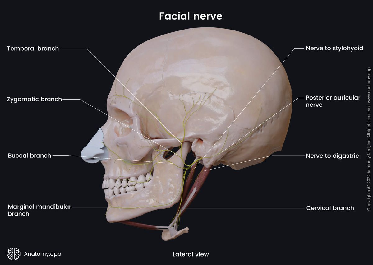
Immediately upon emerging on the outer surface of the skull, the facial nerve turns superiorly, running anteriorly along the outer ear, and it gives rise to three motor branches:
- Posterior auricular nerve - a small motor branch that arises from the facial nerve right below the stylomastoid foramen; it branches into two divisions - auricular branch that innervates the auricularis posterior muscle, and the occipital branch supplying the occipital belly of the occipitofrontalis muscle;
- Digastric branch - it arises from the facial nerve below the stylomastoid foramen and innervates the posterior belly of the digastric muscle;
- Stylohyoid branch - innervates the stylohyoid muscle.
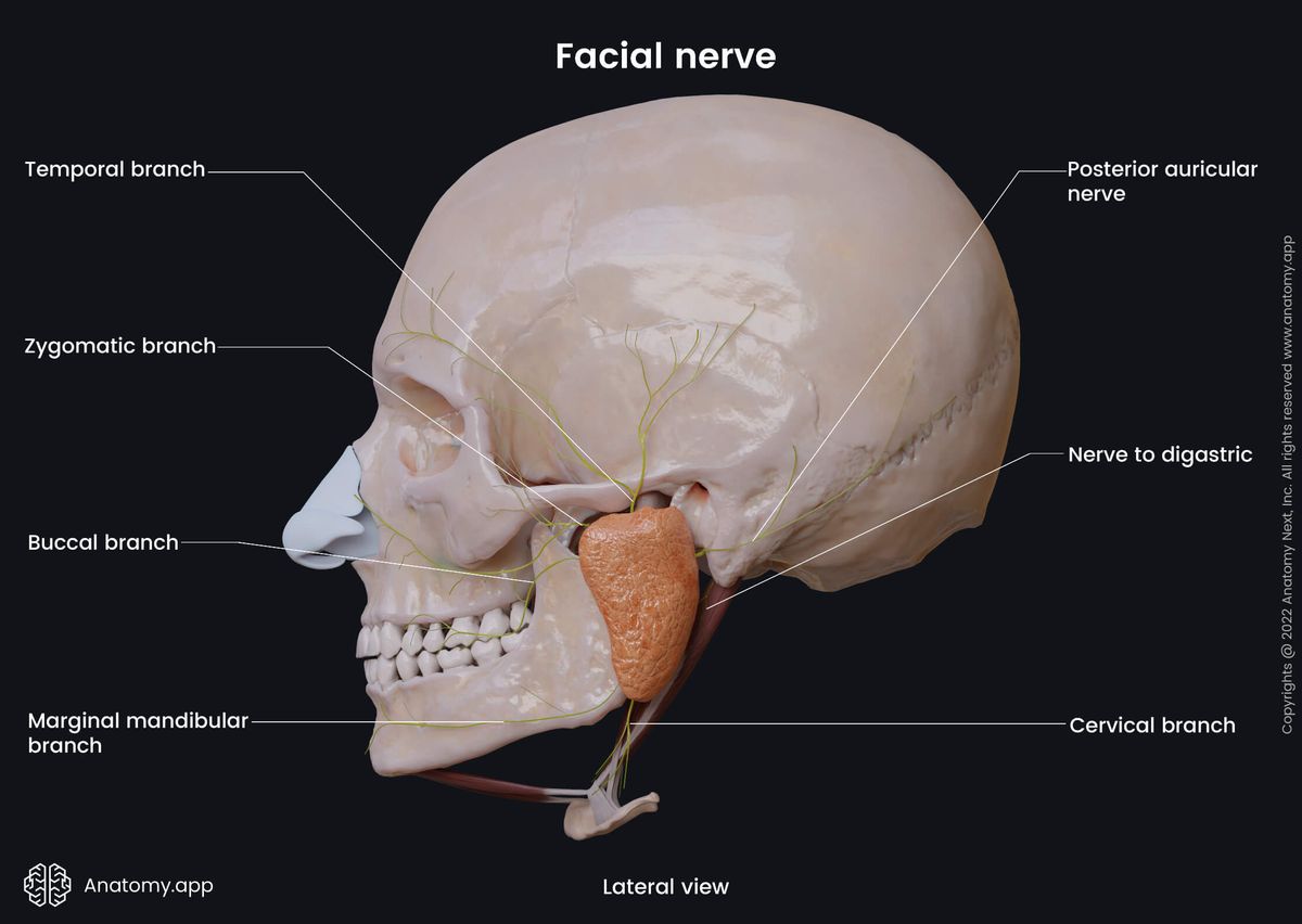
The rest of the motor part (carrying the majority of the motor fibers) of the facial nerve continues its course anteriorly and inferiorly. It divides inside the mass of the parotid gland forming the parotid nerve plexus. (Note: the facial nerve does not participate in innervating the parotid gland, which receives nerve supply from the glossopharyngeal nerve (CN IX).)
Branches from the parotid plexus spread in a fan-like manner to innervate the muscles of facial expression. These branches include the following:
- Temporal branches
- Zygomatic branches
- Buccal branches
- Marginal mandibular branch
- Cervical branch
After arising from the parotid plexus, the temporal branches run ventrally and upwards towards the temporal area and innervate muscles of the auricle - auricularis anterior and auricularis superior - and those muscles of facial expression which are located above the opening of the orbit, such as the frontal belly of occipitofrontalis (also known as the frontalis), orbicularis oculi and corrugator supercilii.
The zygomatic branches pass obliquely, ventrally, and upward, innervating muscles of the lower half of the orbit - orbicularis oculi, zygomaticus major and zygomaticus minor muscles.
The buccal branches leave the parotid plexus in a fan-shaped manner, eventually reaching the area of the mouth and innervating the following muscles of facial expression: zygomaticus major and minor, levator labii superioris, levator anguli oris, risorius, buccinator, orbicularis oris, nasalis and levator labii superioris alaeque nasi.
After leaving the parotid plexus, the marginal mandibular branch runs by the base of the mandible, and innervates muscles of facial expression around the chin - depressor labii inferioris, depressor anguli oris, as well as the mentalis.
Finally, the cervical branch of the facial nerve runs anteriorly beneath the platysma, gives off a branch that passes downward, and joins the cervical cutaneous nerve from the cervical plexus, while other of its branches innervate the platysma.
Intermediate portion of facial nerve
The intermediate portion of the facial nerve (also called the intermediate nerve) is a part of the seventh cranial nerve within the facial canal, which carries parasympathetic and sensory (somatic and special) fibers. Therefore, the intermediate nerve is a mixed nerve. The nerve arises from the facial nerve at the first turn of the facial canal, where the intermediate nerve has its sensory ganglion called the geniculate ganglion.
The sensory fibers arise from pseudounipolar neurons located in the geniculate ganglion. The dendrites of these neurons transmit taste information from the anterior two-thirds of the tongue. The axons of these pseudounipolar neurons travel via the facial nerve to reach the solitary tract nucleus located in the rhomboid fossa below the striae medullares. This nucleus is shared together with the glossopharyngeal nerve (CN IX) and vagus nerve (CN X).
Besides the solitary tract nucleus, this nerve has one more called the superior salivatory nucleus. It is a parasympathetic nucleus located in the rhomboid fossa above the striae medullares, and it innervates the submandibular and sublingual glands. The intermediate nerve divides into two branches right after forming the geniculate ganglion. These branches are called the greater petrosal nerve (parasympathetic) and chorda tympani (parasympathetic and sensory).
Greater petrosal nerve
The greater petrosal nerve carries parasympathetic preganglionic fibers and leaves the facial canal through the hiatus for the greater petrosal nerve. When reaching the geniculate body, the greater petrosal nerve breaks away and travels anterolaterally to exit the superior surface of the temporal bone through the hiatus for the greater petrosal nerve. The nerve emerges on the anterior surface of the petrous part of the temporal bone and crosses it, going in the anteromedial direction.
The greater petrosal nerve goes slightly inferior and passes under the trigeminal cave (a pouch created by the dura mater) towards the foramen lacerum. Then it penetrates the cartilage of the foramen and reaches the external cranial base. In the foramen lacerum, it joins the deep petrosal nerve from the carotid sympathetic plexus, forming the Vidian nerve (also known as the nerve of the pterygoid canal). This nerve innervates the lacrimal gland and the glands and mucous membrane of the nasal cavity and the palate.
The fibers of the greater petrosal nerve originate from the lower part of the pons. The nerve contains not only preganglionic parasympathetic fibers but sympathetic fibers as well. The parasympathetic fibers exit the brainstem as a part of a separate division of the facial nerve known as the intermediate nerve.
Chorda tympani
The chorda tympani is a branch of the facial nerve carrying sensory and parasympathetic preganglionic fibers. It travels almost the entire length of the facial canal and leaves the canal at its descending end part through the canaliculus for chorda tympani, entering the tympanic cavity. In the tympanic cavity, it goes between the malleus and incus.
The nerve leaves the tympanic cavity, emerging on the external cranial base via the petrotympanic fissure. It then reaches the infratemporal fossa and medial to the lateral pterygoid muscle merges with the lingual nerve (a branch of the mandibular division of the trigeminal nerve, CN V3). While going together with the lingual nerve, the fibers of the chorda tympani is distributed to the anterior two-thirds of the sides and dorsum of the tongue and these fibers supply fungiform and foliate papillae.
The sensory - afferent special (gustatory) - fibers of the chorda tympani transmit taste sensations from the lingual papillae of the anterior two-thirds of the tongue via the lingual nerve, while the efferent parasympathetic preganglionic fibers synapse in the submandibular ganglion to provide secretomotor innervation to the submandibular and sublingual glands.
Anatomy.app
Contact information
- For questions regarding business inquiries. Please contact:
- info@anatomy.app