- Anatomical terminology
- Skeletal system
- Skull
- Skeleton of trunk
- Skeleton of upper limb
- Skeleton of lower limb
- Joints
- Muscles
- Heart
- Blood vessels
- Nervous system
- Respiratory system
- Digestive system
- Lymphatic system
- Female reproductive system
- Male reproductive system
- Endocrine glands
- Eye
- Ear
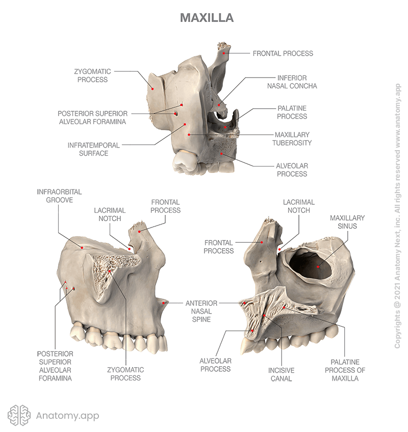
Maxilla
The maxilla (or maxillary bone, upper jaw bone, Latin: maxilla) is a paired bone of the facial skeleton, and it has a body and four processes. The two maxillary bones (maxillae) are fused in the midline by the intermaxillary suture to form the upper jaw.
Parts of maxilla
Each maxilla has five parts, including the body of the maxilla and four processes:
- Frontal process
- Zygomatic process
- Palatine process
- Alveolar process

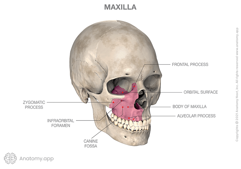
Body of maxilla
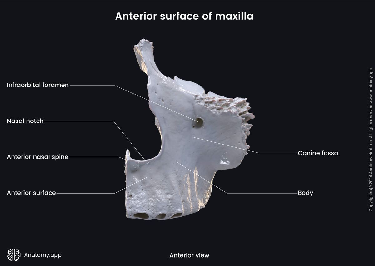
The body of the maxilla is the central portion of the bone housing the maxillary sinus and supporting the four mentioned processes of the maxilla. The body of the maxilla has four surfaces: anterior, orbital, nasal, and infratemporal surfaces.
The anterior surface of the body features the following structures:
- Nasal notch
- Infra-orbital margin
- Infra-orbital foramen
- Canine fossa
The nasal notch is a curved margin of the bony anterior nasal aperture. And the infra-orbital margin is the lower margin of the orbit formed partly by the maxilla, and partly by the zygomatic bone.
The infra-orbital foramen is an opening on the anterior aspect of the maxilla located below the infra-orbital margin. It is the outer opening of the infra-orbital canal that serves as the passage for the infraorbital nerve (a branch of the maxillary nerve, CN V2), accompanied by the infraorbital artery and vein.
The canine fossa is a depressed area on the anterior surface of the body located below the infra-orbital foramen. The canine fossa is the origin site of the levator anguli oris muscle.
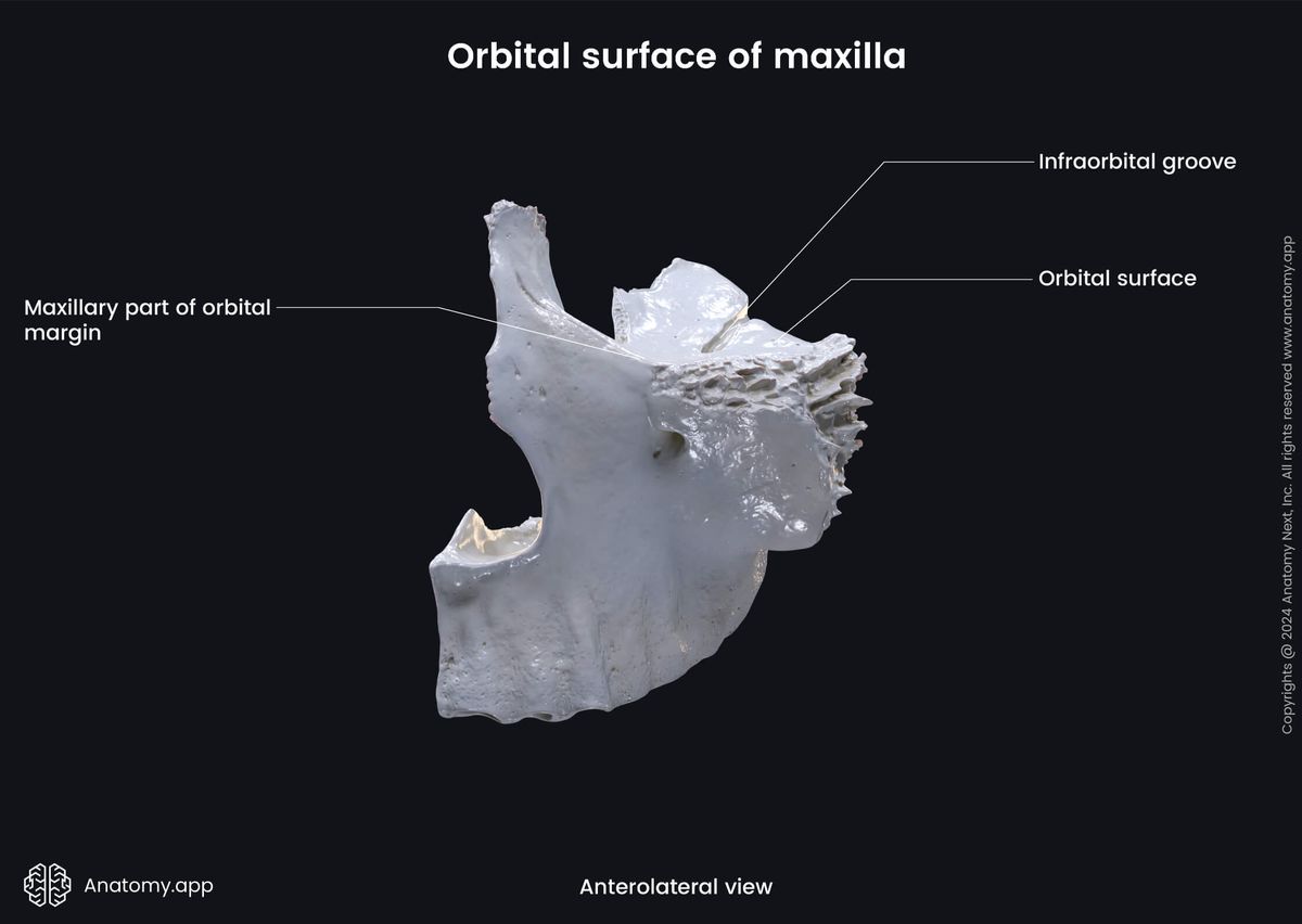
The orbital surface of the body of the maxilla forms most of the floor of the orbit and features the infra-orbital groove leading into the infra-orbital canal. This canal ends with an opening on the anterior surface of the maxilla and serves as the passage for the infraorbital nerve and blood vessels, as mentioned before.
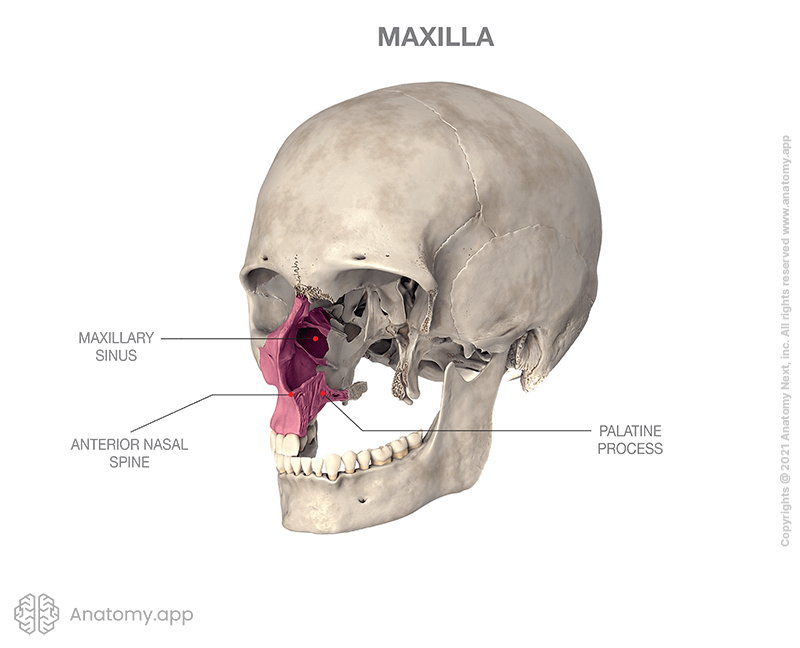
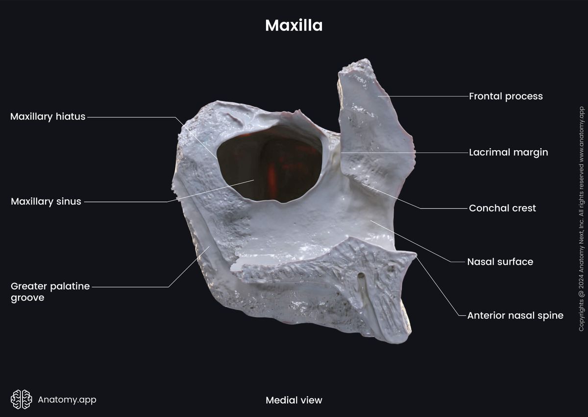
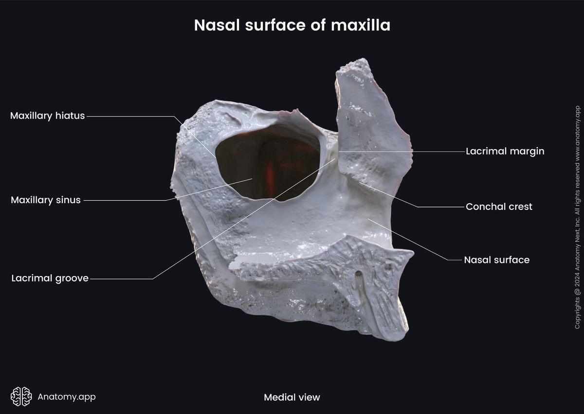
The nasal surface of the body of the maxilla forms part of the lateral wall of the nasal cavity and features a large defect or opening - the maxillary hiatus. The maxillary hiatus is an opening to the maxillary sinus, located below the middle nasal concha. The maxillary sinus is a paranasal sinus, an air-filled cavity located within the body of the maxilla.
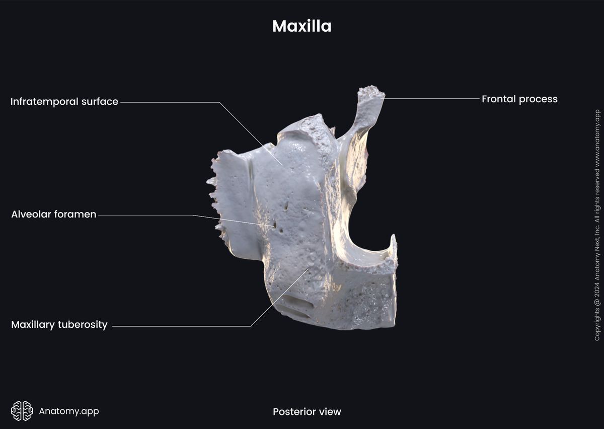
The infratemporal surface of the body of the maxilla features the maxillary tuberosity with alveolar foramina that lead into the alveolar canals. The maxillary tuberosity (or maxillary eminence) is a rounded eminence at the lower part of the infratemporal surface. It is the origin site for a few fibers of the medial pterygoid muscle.
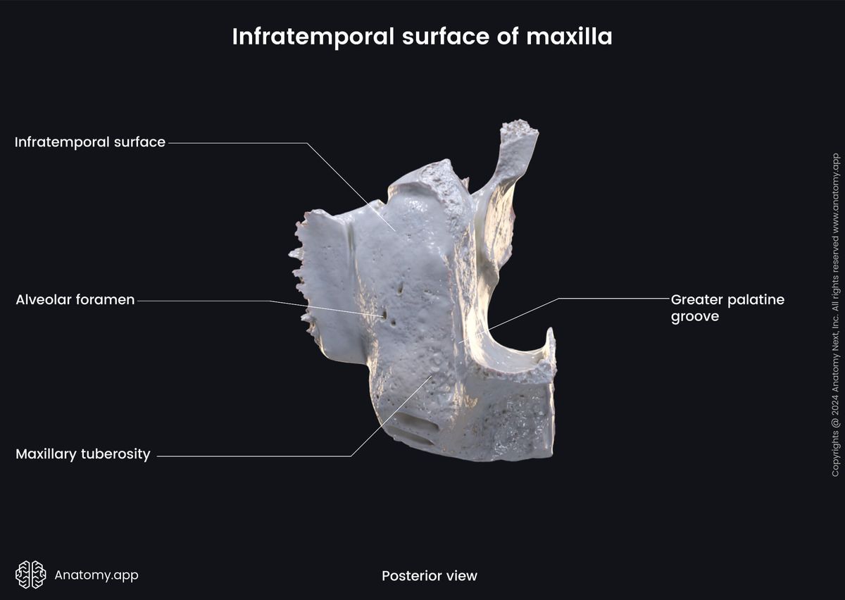
The alveolar foramina are several small openings on the maxillary tuberosity leading into the alveolar canals - several small bony passages - that transmit the posterior superior alveolar nerves (branches of the maxillary nerve) and posterior superior alveolar arteries and veins to the upper teeth.
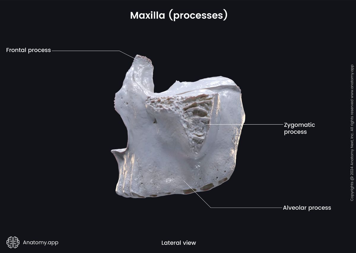
Processes of maxilla
Each maxilla is composed of five parts. Besides the central part known as the body, the maxilla has the following four processes:
- Frontal process
- Zygomatic process
- Palatine process
- Alveolar process
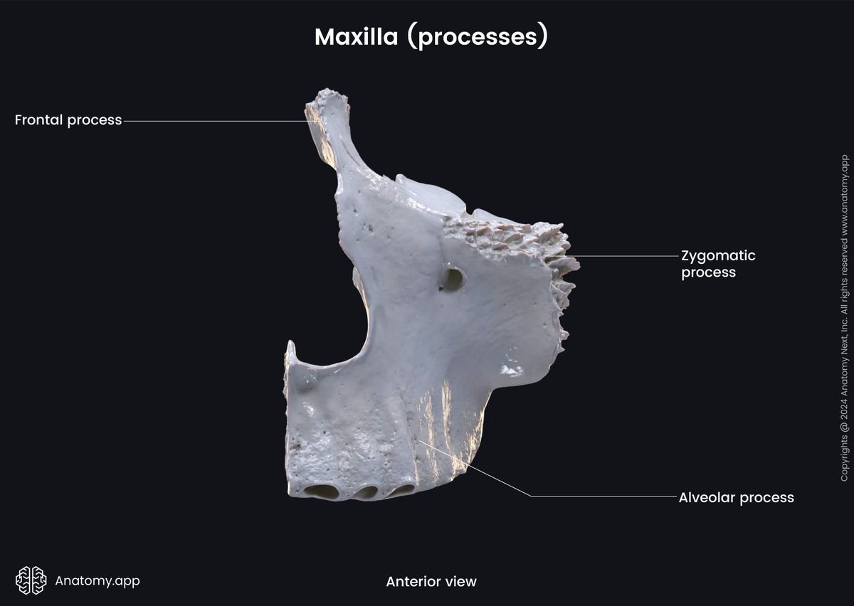
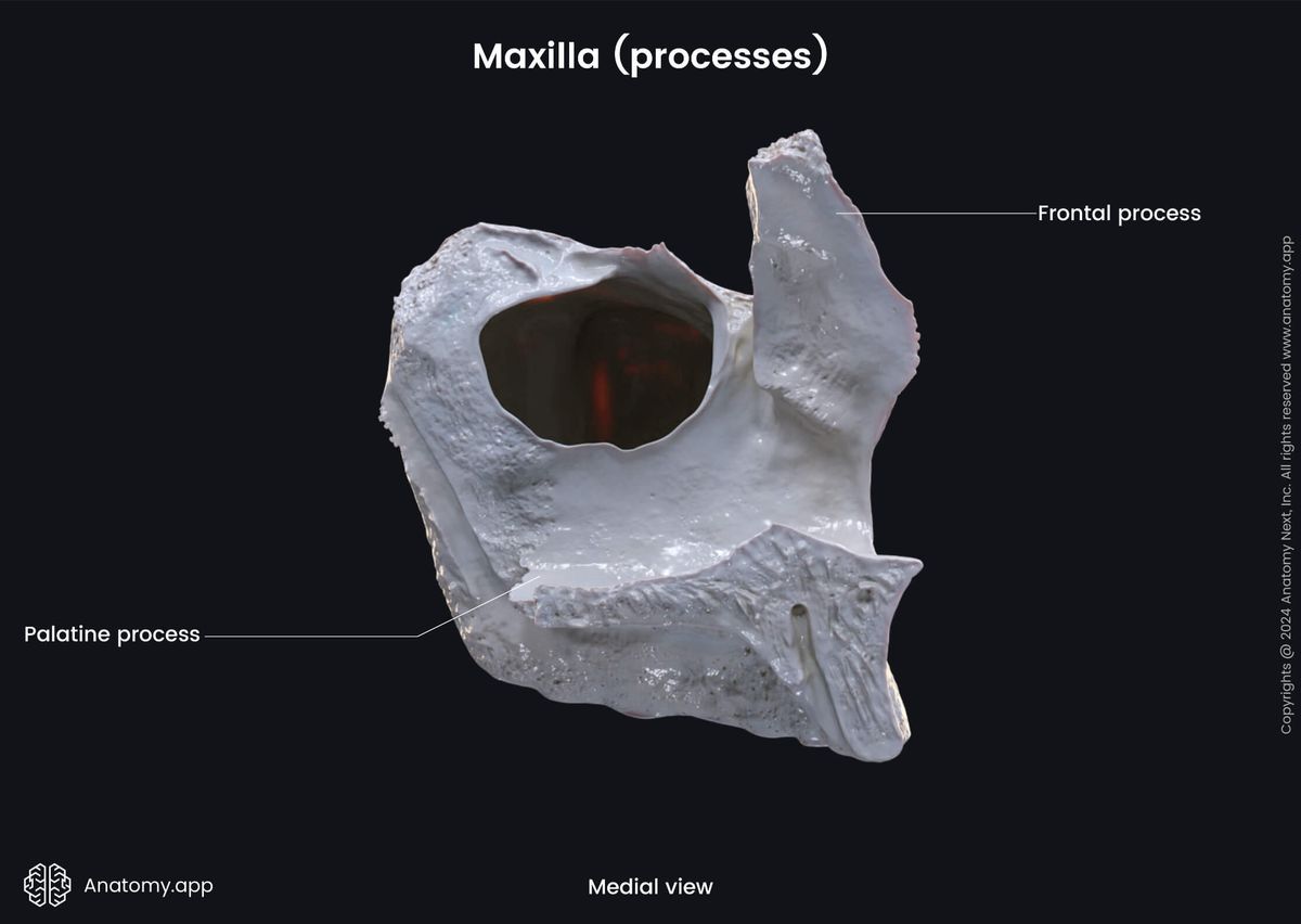
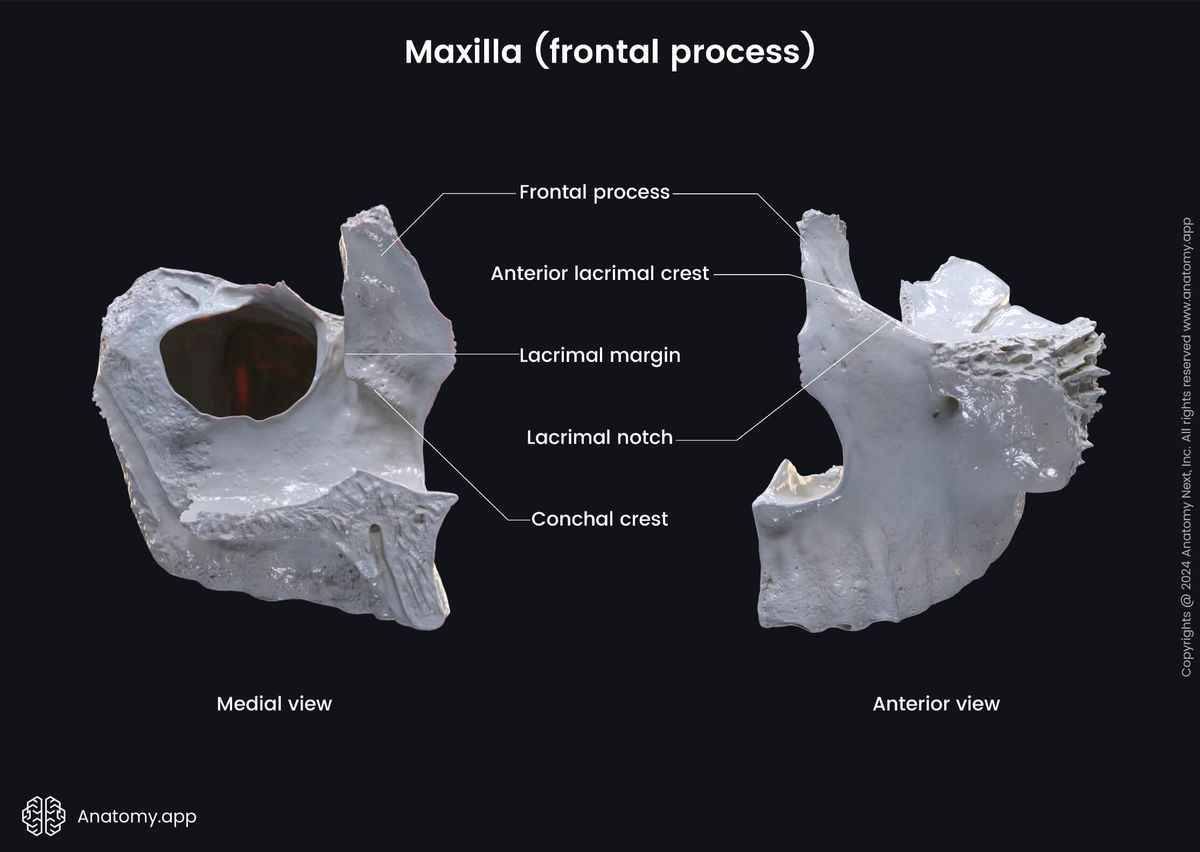
The frontal process is an extension of the maxilla projecting upward, medially and backward for articulation with the frontal bone. The frontal process of the maxilla features the lacrimal groove. Situated on the frontal process of the maxilla and the lateral surface of the lacrimal bone the lacrimal groove participates in forming the nasolacrimal canal. It houses the nasolacrimal duct.
The zygomatic (malar) process of the maxilla is the lateral extension of the bone for articulation with the zygomatic bone. Posterior part of this process is concave and participates in forming the infratemporal fossa. Anteriorly, it forms part of the anterior surface of the maxilla.
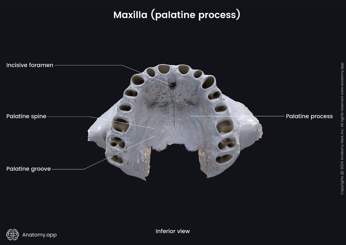
The palatine process of the maxilla is an extension of the maxillary bone shaped as a horizontal plate forming the largest part of the hard palate. The opening of the incisive canal can be found on the palatine process. It starts as a paired canal from the floor of the nasal cavity and unites with the palate in the uniform incisive fossa. From each incisive canal ascends the terminal branch of the greater palatine artery, and descends the nasopalatine nerve.
An alveolar process is a crested process of the upper or lower jaw which houses the teeth. The curved free margin of the alveolar process is called the alveolar arch. The alveolar arch of the maxilla (as the alveolar arch of the mandible) features the following structures:
- Dental alveoli
- Interalveolar septa
- Interradicular septa
- Alveolar yokes
The dental alveoli are sockets in the alveolar process where the roots of the teeth lie. The dental alveoli of the mandible house the roots of the lower teeth, while the dental alveoli of the maxilla - the upper teeth.
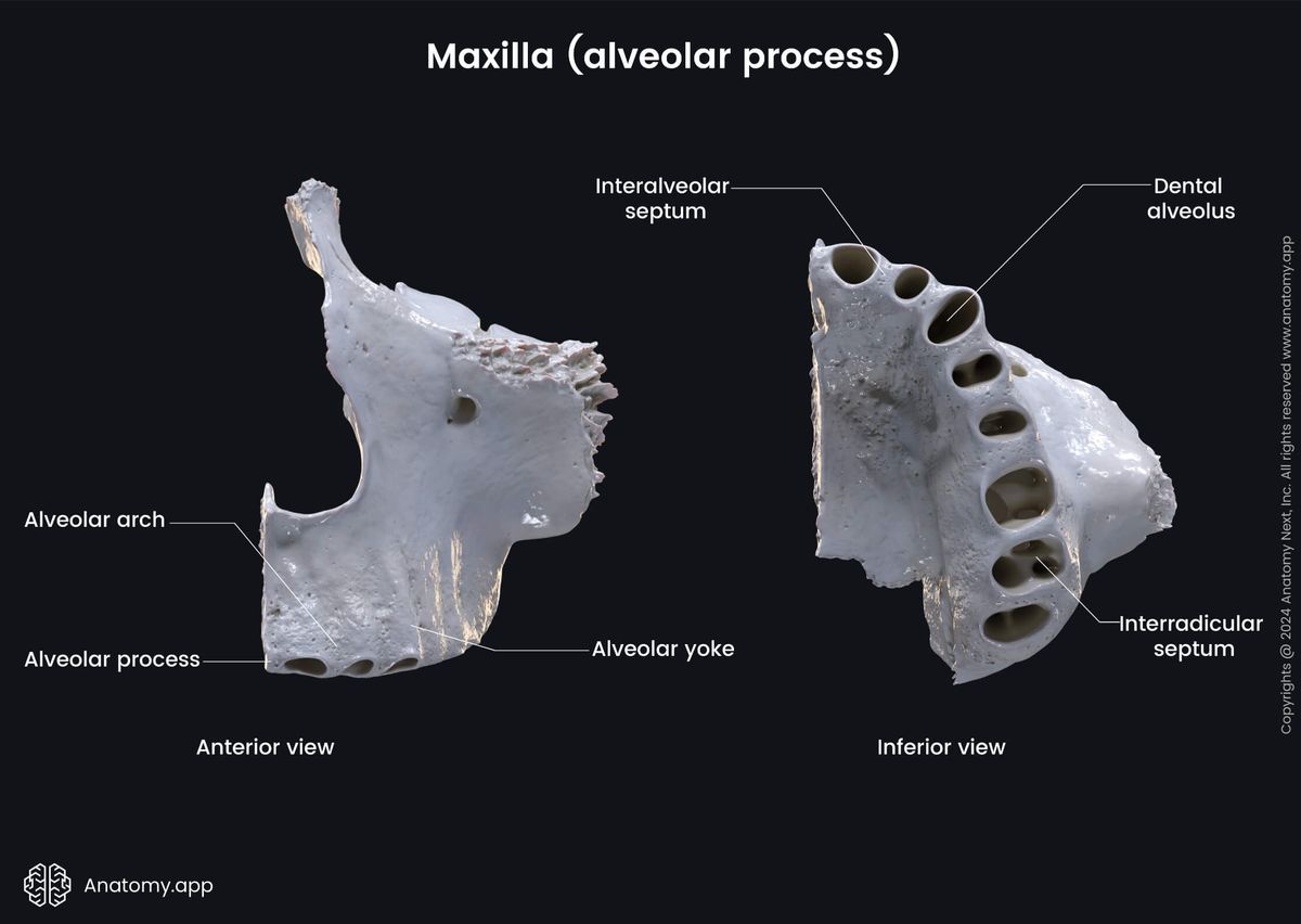
The interalveolar septa are bony ridges between adjacent dental alveoli. But the interradicular septa are bony ridges forming compartments in dental alveoli for the roots of the teeth in both the upper and lower jaw bones.
The alveolar yokes (or juga alveolaria) are eminences on the outer surfaces of the jaw bones produced by the projections of the dental alveoli. They can be seen on alveolar processes of both the maxilla and the mandible.
Anatomy.app
Contact information
- For questions regarding business inquiries. Please contact:
- info@anatomy.app