- Anatomical terminology
- Skeletal system
- Joints
- Muscles
- Heart
- Blood vessels
- Nervous system
- Respiratory system
- Digestive system
- Lymphatic system
- Female reproductive system
- Male reproductive system
- Endocrine glands
- Eye
- Ear
Middle ear
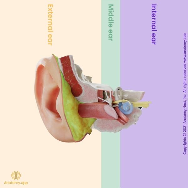
The middle ear (Latin: auris media) includes the tympanic cavity and the auditory tube that connects the tympanic cavity with the pharynx. The middle ear is a quite complex system of openings and canals. Most of this system is located in the temporal bone.
The middle ear is the part of the ear between the tympanic membrane (separating it from the external ear) and the oval window of the inner ear. The tympanic cavity of the middle ear houses three tiny bones, the auditory ossicles, which transfer the vibrations of the tympanic membrane into the waves in the fluid and membranes of the inner ear.
The tympanic cavity is directed medial to the tympanic membrane, but the other part of the middle ear - the epitympanic recess - is located superior to the tympanic membrane. The middle ear is responsible for transferring the compression waves generated in the air into the fluid-membrane waves within the cochlea of the inner ear.
In front of the tympanic cavity is the auditory tube that continues into the pharynx, but behind the tympanic cavity are the mastoid cells of the mastoid process of the temporal bone.
The auditory ossicles work together to receive, amplify and transmit the sound to the inner ear. The ossicles are the malleus, incus, and stapes. The malleus receives vibrations from the tympanic membrane. Afterward, the malleus transmits the vibrations to the incus, which then sends the vibrations to the stapes. The vibrations of the stapes go through the oval window, which results in the movement of fluid within the cochlea. With the help of ossicles, the sound waves are amplified 15-20 times.
Common middle ear disorders
Like the external ear, also the middle ear can experience disorders. Usually, people can have infections or problems with the tympanic membrane.
Acute otitis media
Acute otitis media is an acute infection and inflammation of the middle ear. The condition is characterized by inflammation and edema of the auditory tube mucosa. Due to the different anatomical characteristics of the auditory tube, acute otitis media is more common in children. The acute otitis media is usually seen together with an acute upper respiratory tract infection.
The most commonly seen etiology is Streptococcus pneumoniae and Haemophilus influenzae. During otoscopy, the classical findings are that the tympanic membrane has erythema, bulging that obscures the malleus, thickening with a grayish-white or yellow hue. Also, otalgia and febrility can be present. Treatment of the acute otitis media is antibiotics and analgesics.
Serous otitis media
Serous otitis or serous effusion of the middle ear can start from an upper respiratory tract infection or previously treated acute otitis media. The tympanic membrane usually is retracted, thickened, and shiny. Behind the membrane, a clear yellowish effusion is seen.
Chronic otitis media with effusion
Chronic otitis media with effusion is seen a lot in children. This disorder is characterized by intermittent or chronic hearing impairment, intermittent discomfort, and recurrent infection. A big part of the tympanic membrane is scarred and thickened and can lead to perforation.
Mass lesions in the tympanic membrane
Cholesteatoma is the most commonly seen mass lesion. Cholesteatoma can be whether a defect in the tympanic membrane or a cystic mass. The treatment usually is surgery. Otherwise, the cholesteatoma can grow bigger, invade the mastoid bone and even the inner ear or cranium. Progressive hearing loss is a standard feature.
Another typical lesion is granulomas. Granuloma can result from a cholesteatoma or an old, retained tympanostomy tube. If antibiotics are not helpful, surgery is indicated.
Anatomy.app
Contact information
- For questions regarding business inquiries. Please contact:
- info@anatomy.app