- Anatomical terminology
- Skeletal system
- Skull
- Skeleton of trunk
- Skeleton of upper limb
- Skeleton of lower limb
- Joints
- Muscles
- Heart
- Blood vessels
- Nervous system
- Respiratory system
- Digestive system
- Lymphatic system
- Female reproductive system
- Male reproductive system
- Endocrine glands
- Eye
- Ear
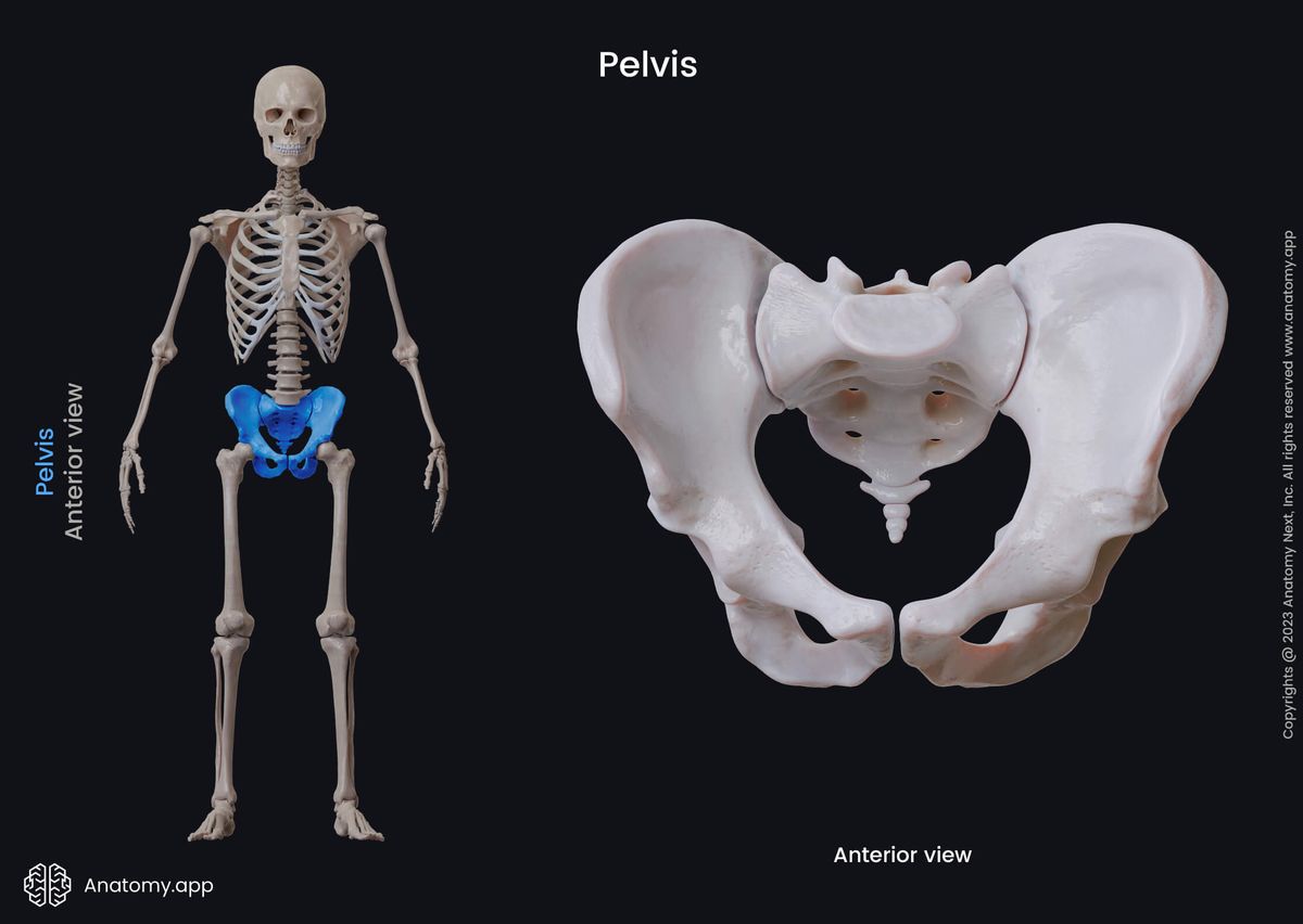
Pelvis
The pelvis (Latin: pelvis) is an anatomical structure located between the abdomen and free lower limbs. The term `pelvis` refers to the pelvic skeleton known as the pelvic girdle. The pelvic girdle is embedded in the lower part of the trunk, and it connects the axial skeleton with the skeleton of the lower limbs.
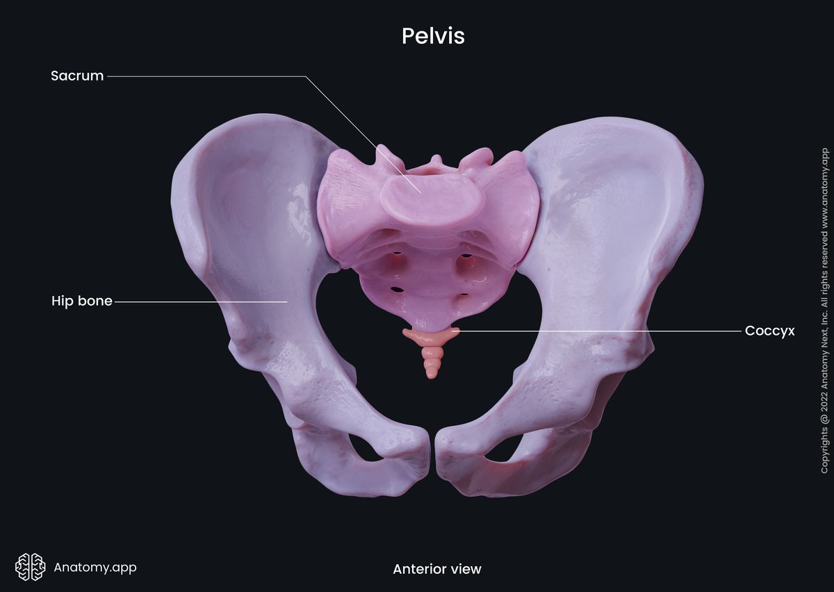
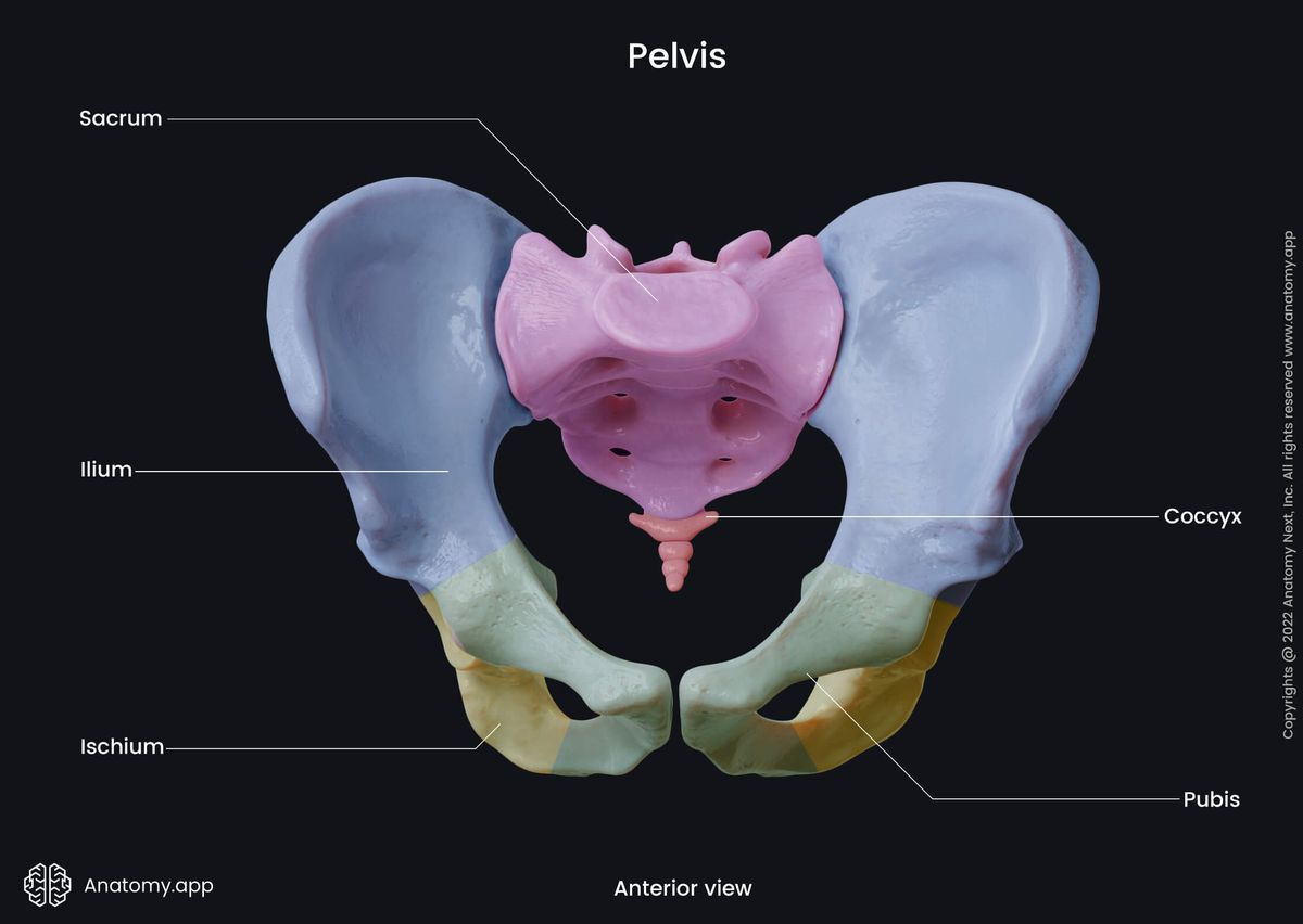
The pelvic skeleton is composed of several bones, including the unpaired sacrum and coccyx and a pair of hip bones. Each hip bone is made of three smaller bones named the ilium, ischium and pubis.
The adjacent bones of the pelvis are connected by four joints, and they are as follows:
- Sacroiliac joints (2) - articulations between the ilium and sacrum;
- Sacrococcygeal symphysis - joint between the sacrum and coccyx;
- Pubic symphysis - articulation between both bodies of the pubis.
Functions of pelvis
The pelvic skeleton is a strong and rigid anatomical structure adapted for several functions in the human body. These functions include the following:
- The pelvis transfers weight from the upper axial skeleton to the lower extremities, especially during the motion.
- It also provides attachment for many muscles and ligaments used in locomotion.
- The pelvis encloses and protects organs located in the abdominal and visceral cavities.
- It also provides natural childbirth when the neonate passes through the birth canal that lies in the pelvic girdle.
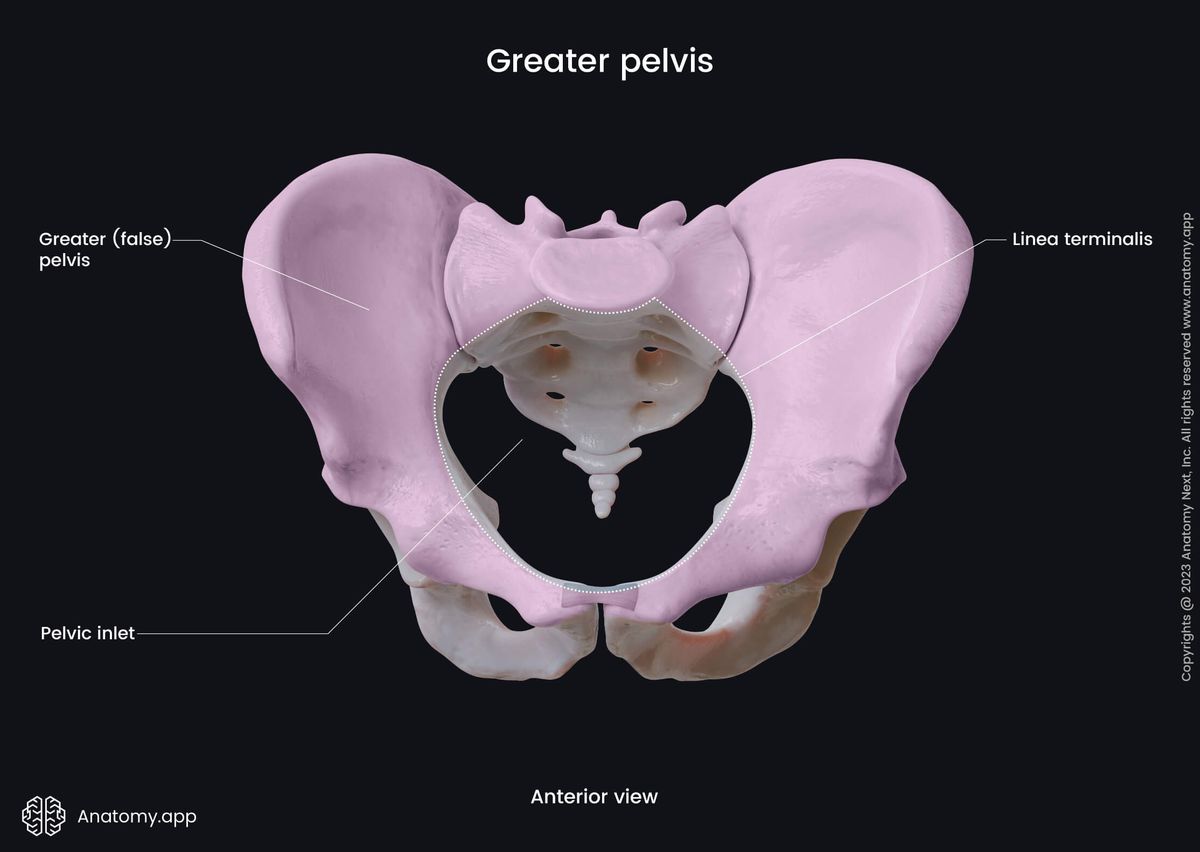
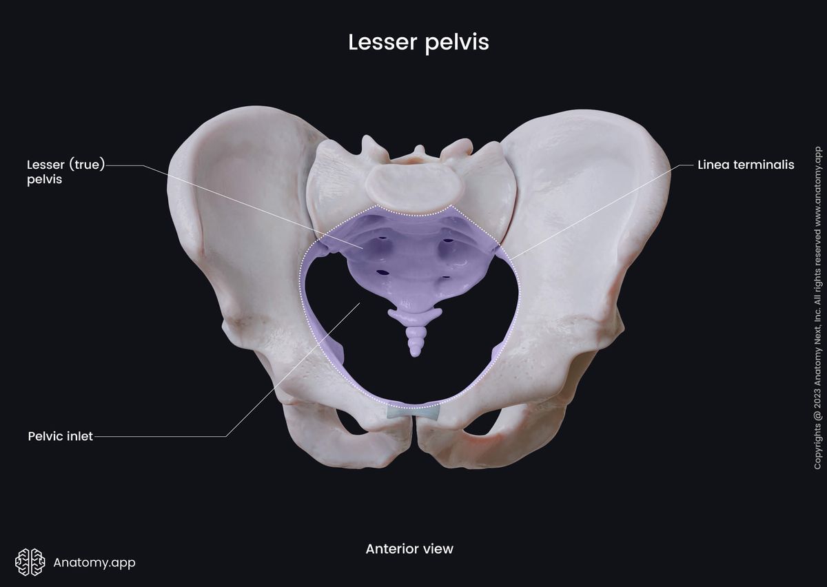
Greater and lesser pelvis
The pelvis is subdivided into two smaller parts - the greater and lesser pelvis - and both parts are separated by the terminal line. The junction between the greater and lesser pelvis is also known as the pelvic inlet.
The greater pelvis, known as the false pelvis, is a space located above the pelvic inlet between the two wings of the ilium. Its primary function is to support the lower abdominal organs, and it has little obstetric relevance.
The lesser pelvis or the true pelvis is a part of the pelvis situated below the pelvic inlet. It encloses the pelvic cavity housing the pelvic organs, including the sigmoid colon, rectum, bladder and several reproductive organs.
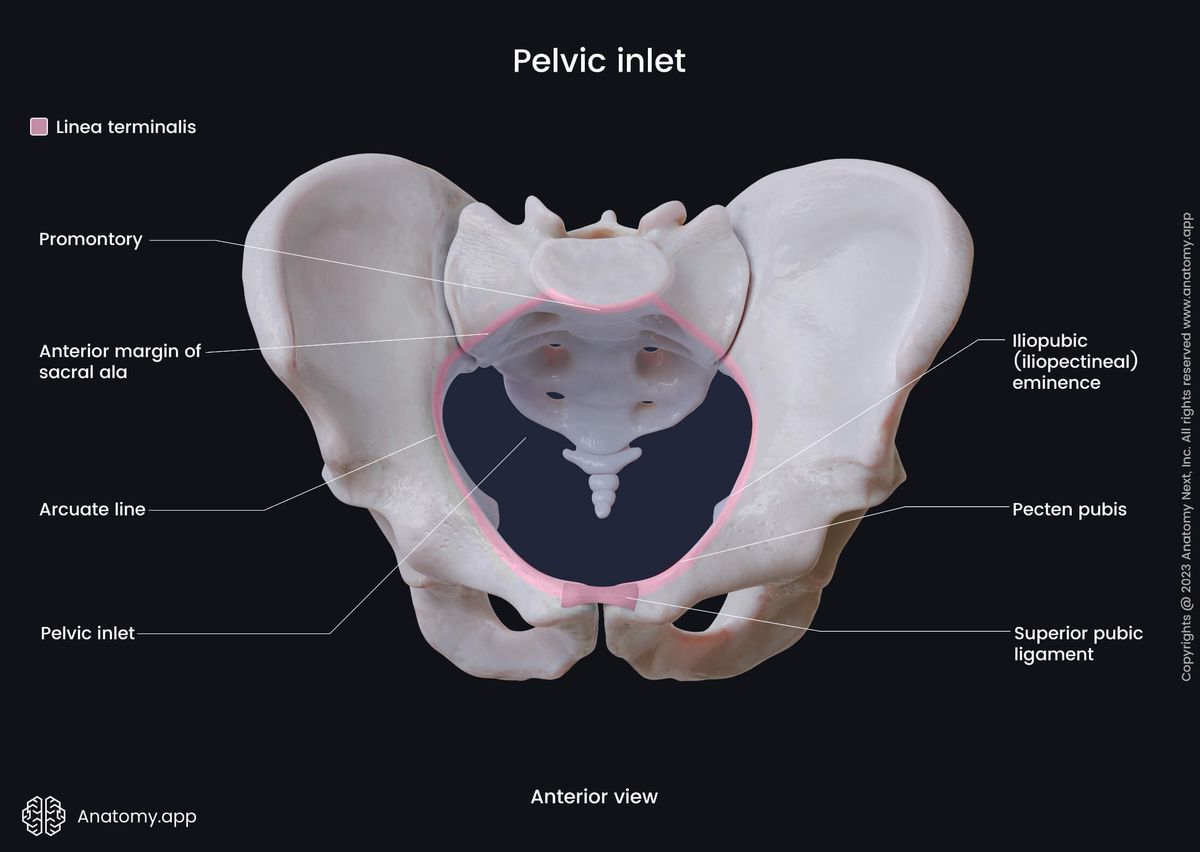
Pelvic inlet
The pelvic inlet marks the junction between the lesser and greater pelvis. The following structures form the borders of the pelvic inlet:
- Posteriorly - anterior sacral promontory and the lower anterior aspects of the wings of the ilium;
- Laterally - arcuate line on the inner surface of the ilium, and the pecten pubis on the superior ramus of the pubis;
- Anteriorly - pubic symphysis and superior pubic ligament.
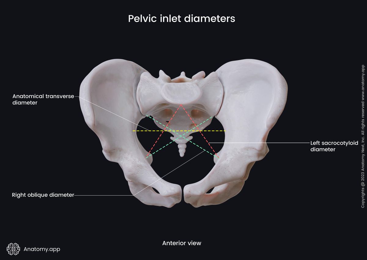
The measurements of the pelvic inlet determine the size and shape of the birth canal. Standard measurements and diameters of the pelvic inlet are as follows:
Anatomical transverse diameter
- Between: the extreme lateral points of the pelvic inlet
- Length: 13 - 14 cm
Oblique diameter
- From: the sacroiliac joint
- To: the iliopubic eminence of the opposite side
- Length: 11.5 - 12.5 cm
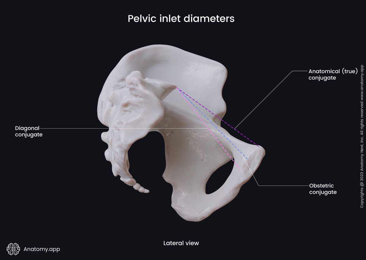
Diagonal conjugate
- From: the inferior pubic ligament
- To: the sacral promontory
- Length: 12.5 - 13 cm
Anatomical conjugate
- From: the superior margin of the pubic symphysis, superior pubic ligament
- To: the sacral promontory
- Length: 11 - 12 cm
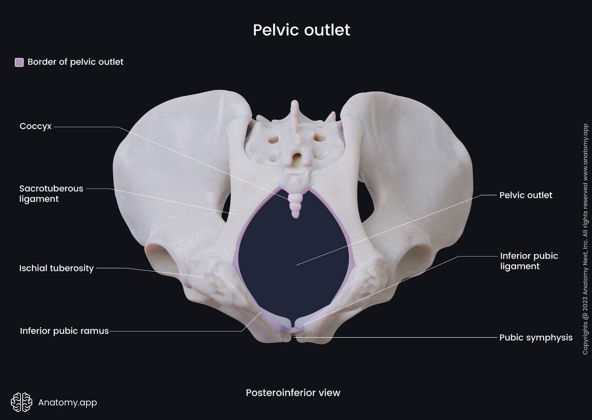
Pelvic outlet
The pelvic outlet, also known as the inferior pelvic aperture, marks the lower aspect of the lesser pelvis, and its shape is very irregular. The boundaries of the pelvic outlet are formed by the coccyx, sacrotuberous ligaments, ischial tuberosities, both ramus of the ischium, both inferior ramus of the pubis and pubic symphysis.
Standard diameters and measurements of the pelvic outlet include the following:
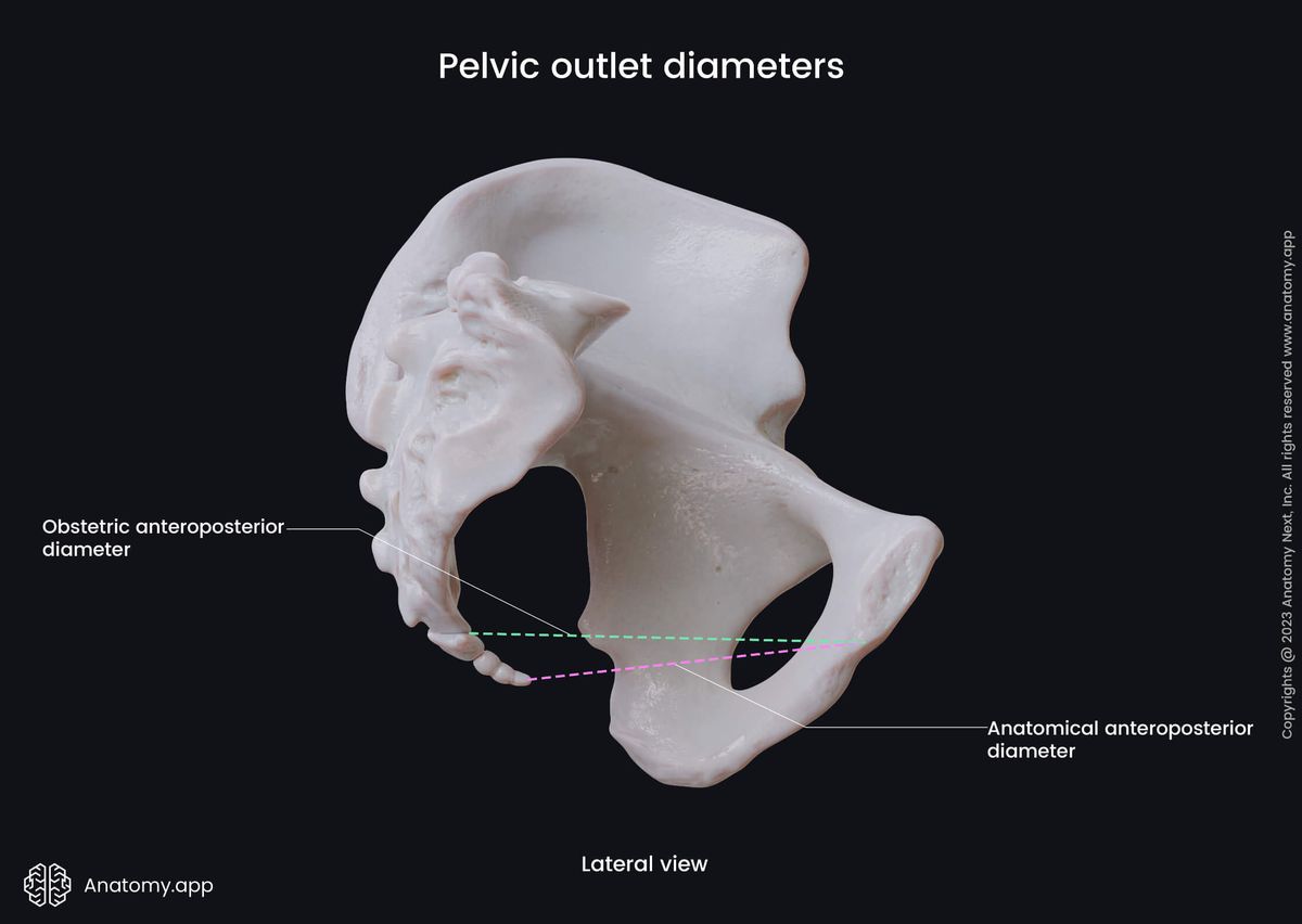
Anatomical anteroposterior diameter
- From: the lower border of the pubic symphysis
- To: the tip of the coccyx
- Length: 9.5 - 12.5 cm (this diameter can significantly vary due to the mobility of the coccyx)
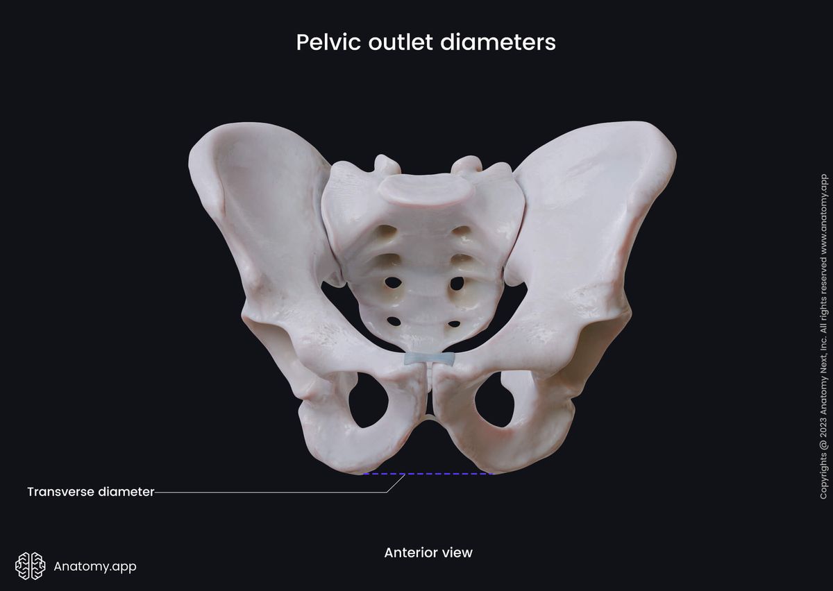
Transverse diameter
- Between: the ischial tuberosities
- Length: 8 - 11 cm
In males, all these diameters are slightly shorter than females, as males usually have narrower and smaller sizes of the pelvis. The diameters and measurements of the female pelvis are important in pelvimetry that assesses the size of a woman's pelvis to predict whether the woman will be able to give natural childbirth. All diameters increase during labor to make the delivery of the fetus easier.
Anatomy.app
Contact information
- For questions regarding business inquiries. Please contact:
- info@anatomy.app