- Anatomical terminology
- Skeletal system
- Joints
- Muscles
- Heart
- Blood vessels
- Nervous system
- Respiratory system
- Digestive system
- Lymphatic system
- Female reproductive system
- Male reproductive system
- Endocrine glands
- Eye
- Ear
Tympanic cavity
The tympanic cavity (Latin: cavitas tympani) is a narrow, irregular space located in the petrosal part of the temporal bone and situated between the external and the internal ear. The tympanic cavity houses three ossicles that provide conduction and amplification of sound vibrations from the tympanic membrane to the inner ear.
Structure of the tympanic cavity
The tympanic cavity has a vertical diameter of around 18 mm, an anteroposterior diameter of about 10 mm, and a transverse diameter of 3 to 5 mm. The tympanic cavity is lined by mucosa with a cylindrical or layered cubic epithelium inter spread with ciliated cells. The epithelium changes to the pseudo-stratified ciliated epithelium, as it is through the entire length of the auditory tube. Apart from the mucosa, the tympanic cavity houses auditory ossicles, two muscles of auditory ossicles, and air.
The tympanic cavity can be divided into three parts - epitympanum (or attic space), mesotympanum, and hypotympanum.
The borders forming the walls of the tympanic cavity are as following:
- The tegmental wall (roof) is formed by the tegmen tympani (squamous and petrous parts of the temporal bone), and it separates the tympanic cavity from the cranial cavity. This wall is located on the anterior surface of the petrous portion of the temporal bone close to its junction with the squamous part of the temporal bone. The wall prolongs forward to cover in the semicanal for the tensor tympani muscle. The wall's lateral edge corresponds with the remains of the petrosquamous suture.
- The jugular wall (floor) is a narrow wall formed by a bone that separates the middle ear from the internal jugular vein. Located above the jugular fossa.
- The membranous wall (lateral wall) is formed mainly by the tympanic membrane (partly by the ring of the bone into which the membrane is inserted) and the bony wall of the epitympanic recess in which auditory ossicles are located. This ring of the bone is not complete in its upper part, thus forming a notch. Near the notch are three openings - anterior chordae, posterior chordae, and the petrotympanic fissure.
- The labyrinthine wall (medial wall) separates the middle ear from the inner ear; it includes the promontory of the labyrinthine wall and the oval and round windows. Both windows lead to the internal ear. The oval window is closed with the basis of the stapes, while the round window is completed with the secondary tympanic membrane. The vibrations of this membrane allow the fluid of the cochlea to move. The promontorium is the projection of the first coil of the cochlea, and its groove acts as a pathway for the tympanic nerve. In this wall also the prominence of the facial canal is located. The prominence has one horizontal and vertical part, with the facial nerve going through the horizontal part.
- The carotid wall (anterior wall) separates the tympanic cavity from the carotid canal and artery; it has an opening called the aditus to the mastoid antrum that connects the tympanic cavity to the mastoid cells. The wall also opens the tympanic orifice of the auditory tube. At the upper part of the anterior wall is the orifice of the semicanal for the tensor tympani muscle. They are separated from each other by the septum canalis musculotubarius.
- The mastoid wall (posterior wall) connects the mastoid antrum with mastoid cells. It contains aditus to the mastoid antrum and the pyramidal eminence.
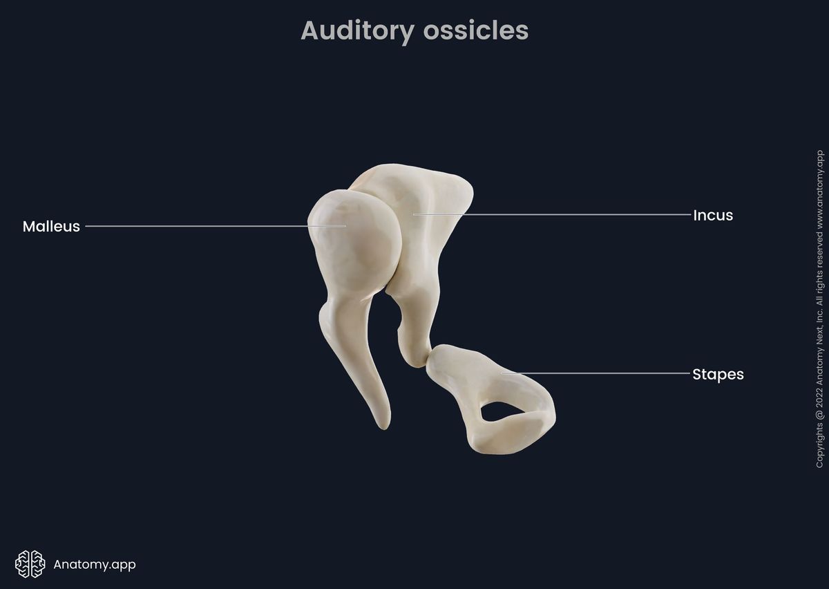
Auditory ossicles
Our ears have three auditory ossicles (Latin: ossicula auditus). They are considered to be the smallest bones in the human body. They work to transmit sounds to the internal ear, directly to the labyrinth. The ossicles articulate with each other through synovial joints. Vibrations are transmitted as a result of the movement of the ossicles after the middle ear's muscles have moved them. Three ossicles are (in the direction from the tympanic membrane to the internal ear):
- Malleus also called hammer
- Incus also called the anvil
- Stapes also called stirrup
One thing that these bones lack compared to the other bones is osteogenic periosteum. There are junctures present between the auditory ossicles:
- Incudomalleolar joint - saddle-shaped;
- Incudostapedial joint - flat;
- Tympanostapedial syndesmosis - between the oval window and the base of the stapes.
Thanks to the junctures, the auditory ossicles form an elastic chain that transmits air vibration from the tympanic membrane to the internal ear.
Malleus
The malleus is laterally attached to the tympanic membrane, while medially, the bone articulates with the incus. The malleus has three parts:
- The head of the malleus - with saddle-shaped articulation surface and is located in the epitympanic recess. The head of the malleus is connected to the tegmental wall of the tympanic cavity through the superior ligament of the malleus;
- The neck of the malleus - is located inferior to the head and has two processes - lateral and anterior. The anterior process is attached to the anterior wall of the middle ear by the anterior ligament of the malleus, while the lateral process is attached to the medial surface of the tympanic membrane by the lateral ligament of the malleus;
- The handle of the malleus - is an extension that is running inferior from the neck of the malleus. It has grown with the tympanic membrane. By drawing in the membrane, the handle of the malleus creates a belly button-like structure called the umbo.
The malleus receives sound vibrations from the tympanic membrane and transfers those to the incus.
Incus
The incus is located between the malleus and the stapes as a connection between these two bones. The incus has three parts as well:
- The body of the incus - has a saddle-shaped articulation surface that articulates with the head of the malleus through the incudomalleolar joint. The body is found in the epitympanic recess;
- The short limb of the incus - this structure runs posteriorly and is attached to the posterior wall of the tympanic cavity with the help of the posterior ligament of the incus;
- The long limb of the incus - can be found parallel to the handle of the malleus. The long limb ends medially with the lenticular process. The process takes part in articulation with the stapes via the incudostapedial joint.
Stapes
The stapes is articulating with the incus via the incudostapedial joint on the lateral side, but on the medial side, the bone is attached to the membrane of the oval window on the labyrinth wall of the tympanic cavity. The vibrations that are first caught by the malleus are then carried through the incus to the stapes, which then causes the membrane on the oval window to vibrate. Following this, the sound is then transmitted to the vestibule of the internal ear.
The stapes has the following parts:
- The head of the stapes - articulates with the lenticular process of the long limb of the incus;
- The anterior and posterior limbs of the stapes - attached to the oval base;
- The base of the stapes - fits into the oval window.
Muscles of the auditory ossicles
The middle ear has the smallest skeletal muscles in the human body. Nevertheless, these muscles are essential due to their synergistic actions enabling sound transmission. Apart from the sound transmission, these muscles also protect the internal ear from stimuli that may be too strong by controlling the movements of the ossicles. These two muscles are:
- The tensor tympani muscle - originates from the cartilaginous part of the auditory tube, the greater wing of the sphenoid bone, and the semicanal for tensor tympani. The muscle ends in the superior part of the handle of the malleus. The tensor tympani muscle is responsible for pulling the malleus to the medial side resulting in the tympanic membrane tension and pushing the stapes into the oval window. The muscle is innervated by the mandibular branch (CN V3) of the trigeminal nerve (CN V);
- The stapedius muscle - originates from the inside of the pyramidal eminence of the tympanic cavities mastoid wall. The muscle insertion site is the neck of the stapes. The stapedius muscle pulls the incus to the lateral side, removing the stapes out of the oval window and preventing excessive oscillation. The muscle is innervated by the facial nerve (CN VII).
Vasculature and innervation of the tympanic cavity
Blood supply and venous drainage
The tympanic cavity is supplied by:
- The anterior tympanic artery (a branch of the maxillary artery -> the external carotid artery)
- The posterior tympanic artery (a branch of the stylomastoid artery -> the posterior auricular artery -> the external carotid artery )
- The superior tympanic artery (a branch of the middle meningeal artery -> the maxillary artery)
- The inferior tympanic artery (a branch of the ascending pharyngeal artery -> the external carotid artery)
- Mastoid branch the occipital artery
Venous drainage happens through the tympanic veins into the superior petrosal sinus and the pterygoid venous plexus.
Lymphatic drainage
The lymph from the tympanic cavity is drained into the retroauricular cervical lymph nodes.
Innervation
The mucosal side of the tympanic membrane is innervated by tympanic branches of the glossopharyngeal nerve (CN IX). The same nerve is responsible for innervating the tympanic cavity.
Anatomy.app
Contact information
- For questions regarding business inquiries. Please contact:
- info@anatomy.app